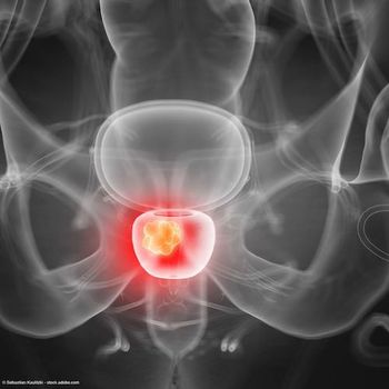
Pivotal data for PSMA PET imaging agent 18F-DCFPyL shared as FDA weighs approval in prostate cancer
The FDA is scheduled to make a decision on a new drug application for 18F-DCFPyL on or before May 28, 2021.
Data from the pivotal phase 3 CONDOR study of 18F-DCFPyL in patients with biochemically recurrent prostate cancer were shared at the 2021 Genitourinary Cancers Symposium as the FDA prepares to make a decision on a new drug application (NDA) for the PSMA PET imaging agent.1
The multicenter phase 3 CONDOR study (NCT03739684) enrolled men with rising PSA after definitive therapy and negative or equivocal standard-of-care imaging. Patients were required to have a PSA level ≥0.2 if they had undergone radical prostatectomy (RP) or a PSA level ≥2.0 if they were treated with radiation therapy or cryotherapy.
The radiolabeled small molecule 18F-DCFPyL targets the extracellular domain of PSMA. Imaging is supposed to begin 1 to 2 hours following intravenous administration of 18F-DCFPyL as bolus injection.
The primary end point of the study was correct localization rate (CLR), defined as percentage of patients with a 1:1 correspondence between at least 1 lesion identified by Peel–PET/CT and the composite standard of truth (SOT; N = 132: pathology, correlative imaging, or PSA response). Peel scans were read by 3 blinded independent central readers. The key secondary end point was the percentage of patients in whom 18F-DCFPyL PET/CT led to a change in intended treatment.
Overall, there were 208 evaluable patients, about 85% of whom underwent RP, either alone or with radiation. Median PSA level of the cohort was 0.8 ng/mL, and 68.8% had a PSA level <2.0 ng/mL. Some 27.9% had received at least 1 prior systemic therapy. Detection of disease as manifested by a positive 18F-DCFPyL–PET/CT scan was 65.9%, 59.6%, and 59.1% by the 3 readers.
The prespecified criterion for CLR success was for the lower limit of the 95% CI to exceed 20% for at least 2 of the 3 readers. For every reader, the lower bound of the 95% CI for the CLR was well in excess of the 20% benchmark, meeting the primary end point of the study.
The CLRs were 85.6% (95% CI, 78.8%-92.3%), 87.0% (95% CI, 80.4%-93.6%), and 84.8% (95% CI, 77.8%-91.9%) by the 3 readers.
Positive predictive value was also high, with a range of 88.7% to 90.7% across all 3 readers. Further, the high CLR and PPV were maintained across all 3 categories of SOT. For histopathology (n = 31) the CLR ranges were 78.6 to 82.8% and the PPV ranges were 92.9% to 93.3%. For correlative imaging (n = 100) the ranges were 86.1% to 88.6% and 87.% to 89.5%, respectively. The ranges were both 100% for PSA response.
“PSMA-targeted 18F-DCFPyL-PET/CT detected and localized metastatic lesions with high CLR and PPV regardless of which criterion defined CLR that was used, in men with BCR who had negative or equivocal baseline standard imaging,” first study author Frederic Pouliot, MD, PhD, assistant professor, department of Surgery, Faculty of Medicine, Université Lavaland, Quebec, Canada, and coauthors wrote.
Regarding the secondary end point of treatment change, as measured through pre- and post-PET questionnaires, 63.9% of evaluable patients had a change in intended management due to 18F-DCFPyL-PET/CT. Of these changes, 78.6% were triggered by positive 18F-DCFPyL-PET/CT findings and 21.4% were caused by negative findings.
The changes included 58 patients switching from salvage local therapy to systemic therapy, 49 patients changing from observation to initiating therapy, 43 patients switching from noncurative systemic therapy to salvage local therapy, and 9 patients scaling back from planned treatment to observation.
The NDA for 18F-DCFPyL-PET/CT being considered by the FDA also includes supportive data from the phase 2/3 OSPREY trial, in which 18F-DCFPyL-PET/CT was assessed in 2 patient cohorts.2 Cohort A included men with high-risk, locally advanced prostate cancer, and the researchers assessed the capacity of 18F-DCFPyL-PET/CT to detect prostate cancer in pelvic lymph nodes. Cohort B comprised patients with metastatic or recurrent disease and the researchers examined the performance of 18F-DCFPyL-PET/CT in detecting distant metastases.
In cohort A, results for 18F-DCFPyL-PET/CT in detecting disease in pelvic lymph nodes showed a specificity of 96% to 99%, a sensitivity of 31% to 42%, and a PPV of 78% to 91%. The sensitivity and PPV rates for detecting metastatic lesions in cohort B were 93% to 99% and 81% to 88%, respectively.
References
1. Pouliot F, Gorin MA, Rowe SP, et al. PSMA-targeted imaging with 18F-DCFPyL-PET/CT in patients (pts) with biochemically recurrent prostate cancer (PCa): A phase III study (CONDOR)—A subanalysis of correct localization rate (CLR) and positive predictive value (PPV) by standard of truth. J Clin Oncol. 2021;39(suppl 6):33. doi: 10.1200/JCO.2021.39.6_suppl.33
2. PDUFA action date of May 28, 2021 assigned by U.S. Food and Drug Administration. Published online December 9, 2020. https://bit.ly/2KtYerG. Accessed February 13, 2021.
Newsletter
Stay current with the latest urology news and practice-changing insights — sign up now for the essential updates every urologist needs.


