Opinion
Article
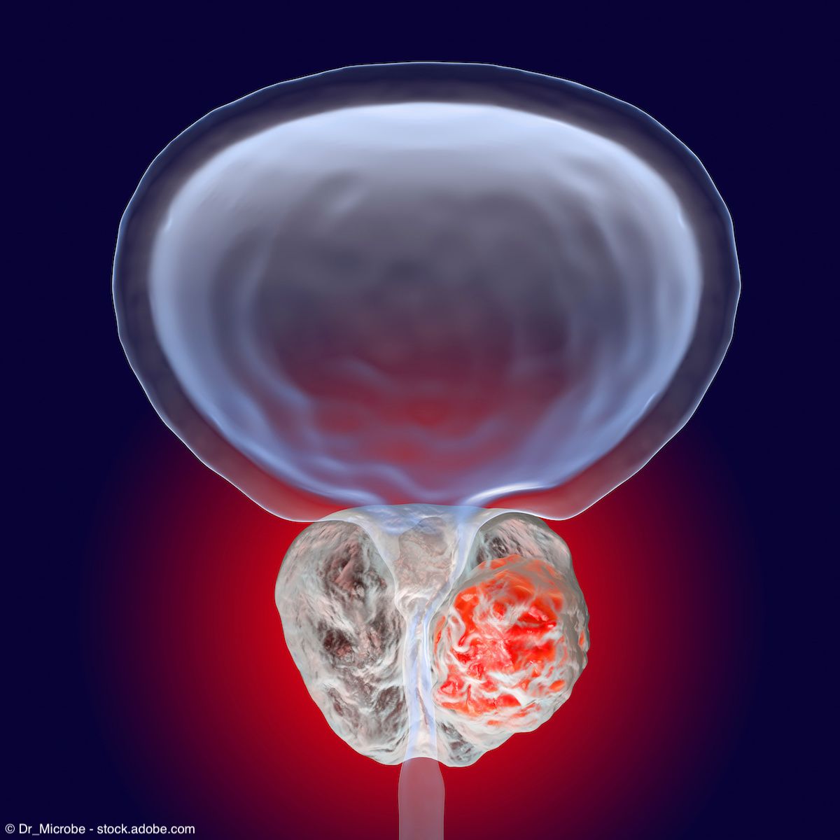
PSMA PET-CT Imaging: Impact on Real-World Diagnosis and Management of Prostate Cancer
Author(s):
In this companion article, Jaideep S. Sohi, MD, shares insights on PSMA-PET imaging’s impact on real-world diagnosis and management of prostate cancer.
Prostate-specific membrane antigen (PSMA)-directed PET-CT imaging is impacting diagnosis, staging, and treatment decisions for patients with prostate cancer. Jaideep S. Sohi, MD, founder and president of American Molecular Imaging and clinical faculty member at Northwestern University School of Medicine, offers his perspective on how this imaging is impacting real-world practice.
Urology Times®: When would you recommend PSMA PET-CT imaging for initial diagnosis and/or restaging?
Jaideep S. Sohi, MD: Until now, we’ve used what we call conventional imaging, which is bone scan and CT imaging for staging and restaging patients. Both are wonderful modalities but come with their own challenges. For example, [with a] bone scan, you need the PSA to be quite high before it demonstrates meaningful findings, at which time the patient may have limited options for treatment.
A CT scan is also a wonderful modality, and we use it on a daily basis. [There are] a couple of challenges with CT scans. Number 1 is that often we’re only including the abdomen and pelvis in the field of view, so [we may be] potentially missing additional sites....And second, with prostrate carcinoma, a significant majority of the lymph nodes that are involved are not enlarged by CT criteria. In other words, we may see them on CT, but it’s difficult to [determine whether] they are malignant.
This is where PSMA PET comes in for initial staging, as opposed to using CT and bone scan. We now have a modality that we can utilize to evaluate the patient’s whole body, including bones and soft tissue. We can also use the same modality in patients who have been treated in the past and are coming back with biochemical recurrence.
Urology Times: Can you briefly discuss the available PSMA PET tracer options and their impact in terms of accurately diagnosing and staging prostate cancer?
Jaideep S. Sohi, MD: Today we have multiple PSMA tracers for evaluation of patients with prostate carcinoma. They are either labeled with gallium 68 or with F18 [fluorine 18]. In our clinical experience, as well as the data that’s out there, they’ve demonstrated similar efficacy and effectiveness in evaluating patients with prostate carcinoma. So, from a real-world perspective, there have been uniform results from these modalities.
In terms of similarities, there are many. For example, the biodistribution of the tracers is very similar. One salient difference is that you may see more background hepatic uptake with [an] F18-based tracer as opposed to gallium 68, but otherwise, the biodistribution is quite similar. There may be more cases of nonspecific uptake in the bones and soft tissues, such as ganglia, with F18 [tracers] compared to that seen with the gallium 68 [tracer]. That can sometimes confound the findings and has the potential of impacting patient management because, as we know, prostrate carcinoma spreads to the bones and it’s critical that we provide [a diagnosis that is] as accurate as possible and interpretation of the findings linked to the bones.
Urology Times: Can you share any tips for interpreting results?
Jaideep S. Sohi, MD: I teach my colleagues, residents, and fellows that you should develop your own protocol and workflow [regarding] how you evaluate these images and keep it consistent. So, for example, we look at the MIP or maximum intensity projection image as a first step and look at the PET image alone in coronal, sagittal, and axial planes, followed by looking at fused images. That gives us a nice flavor for what to expect when we look at and evaluate the study.
With regard to false positives, the most common sites are going to be the bones because of multiple ideologies like posttraumatic changes, fibrous dysplasia, and others, as well as the ganglia, which can sometimes mimic lymph nodes. It can be challenging to resolve whether they indicate the presence of a lymph node or a benign ganglia.
Given that we may potentially see a higher incidence of these false positives or nonspecific uptake with F18 compared to gallium, that’s something that you should keep in mind when reading those studies.
Urology Times: When do you recommend reimaging? How often should imaging be done? At what PSA level?
Jaideep S. Sohi, MD: It depends on the patient’s clinical condition. We [use] PSMA PET imaging when there’s a change in the clinical scenario for the patient or the PSA starts to rise. We do have a definition of biochemical recurrence, which means a PSA of 2.0 or higher measured at 2 different time points for patients who have undergone prior radiation therapy and a PSA of 0.2 or higher measured at 2 different points for patients who have undergone prior prostatectomy. We use these as markers to perform subsequent PSMA PET imaging.
Urology Times: What is the difference in the half-lives of gallium 68 and F18 tracers, and what does this mean in real-world practice in terms of scheduling and preparing for imaging? How do you choose the right tracer?
Jaideep S. Sohi, MD: Gallium 68, as the name implies, has a half-life of 68 minutes, and F18 has a half-life of approximately 110 minutes. With the shorter half-life of gallium, you may have a narrower window in which to scan the patient. And with [the] longer half-life of F18, you may have a greater window in which to scan the patient in terms of scheduling and preparation, so it gives you a little bit more flexibility.
[Specifically], gallium 68 is extracted by a generator and F18 is produced by cyclotron. There are some differences in these 2 techniques. Cyclotrons tend to be more costly [and] introduce a level of complexity. There’s also limited availability of cyclotrons. There are greater energy requirements for deploying cyclotrons and there [are] also additional radiation safety considerations, so it can be a little complex to utilize cyclotrons.
On the other hand, generators are [much] more readily available and we’re able to dilute the desired product multiple times so it can help us with regard to flexibility and being able to acquire the tracer in a more convenient time frame.
With cyclotrons, you’re on a schedule, so it’s sometimes [difficult] to be flexible and pivot to provide a last-minute dose. With on-site or locally available generators, it can be easier to provide that tracer at a last-minute notice. So, for example, if there is a patient who has rescheduled or canceled and there’s an open slot, then from a logistical perspective, it can be easier to provide a generator-extracted product as opposed to a cyclotron[-generated tracer] because of that more complex and lengthy procedure.
It really comes down to which tracer is more readily available when we need to pivot and be flexible with regard to patient cancellations, rescheduling, or being late. Many times patients are coming from out of town, especially in the larger metro areas, where they may not have access to these specialized tracers in their own community. They may have a follow-up appointment with their referring physician [on] the same day. So, it becomes crucial that we are able to scan them on [that] same day. Patients [also] may be debilitated and have challenges with transportation. So, these are things that we have to keep in mind when we’re looking at which tracer to use.
I think PSMA [PET-CT imaging] is really a game changer for patients with prostate carcinoma. If we have access to one or the other, I would choose the one that we have available because they generally work in a similar fashion. Our preference tends to be for [a] gallium-based tracer because of the points raised earlier with regard to increase [in] nonspecific uptake in the bones and soft tissue with F18. But, at the end of the day, if we have only 1 available, we’ll use [the accessible] tracer because these are really making a dramatic change in patient care.


Newsletter
Stay current with the latest urology news and practice-changing insights — sign up now for the essential updates every urologist needs.