
|Articles|June 17, 2021
Update further validates piflufolastat F 18 effectiveness in prostate cancer
Author(s)Jason M. Broderick
Piflufolastat F 18 effectively detected and pinpointed metastatic lesions with high positive predictive value, regardless of anatomic region.
Advertisement
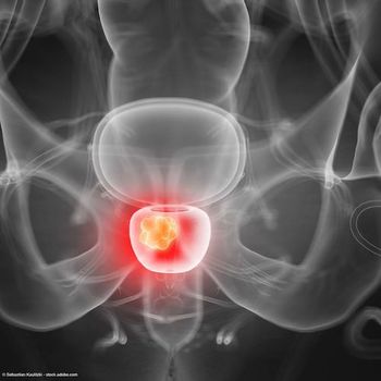
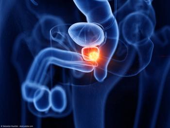
Among patients with biochemically recurrent prostate cancer and negative/equivocal imaging at baseline, piflufolastat F 18 (18F-DCFPyL-PET/CT; Pylarify) effectively detected and pinpointed metastatic lesions with high positive predictive value (PPV), according to an update from the pivotal phase 3 CONDOR trial presented at the 2021 Society of Nuclear Medicine and Molecular Imaging (SNMMI) Annual Meeting.1,2
The researchers noted that PPVs with piflufolastat F 18 were higher in extra-pelvic lymph nodes and bone versus viscera/soft tissue regions. Findings from the CONDOR trial previously supported the
The multicenter phase 3 trial enrolled men with rising PSA after definitive therapy and negative or equivocal standard-of-care imaging. Patients were required to have a PSA level ≥0.2 if they had undergone radical prostatectomy (RP) or a PSA level ≥2.0 if they were treated with radiation therapy or cryotherapy.
The primary end point was correct localization rate (CLR), defined as percentage of patients with a 1:1 correspondence between at least 1 lesion identified by PyL–PET/CT and the composite standard of truth (pathology, correlative imaging, or PSA response). PyL scans were read by 3 blinded independent central readers.
Overall, there were 208 evaluable patients, about 85% of whom underwent RP, either alone or with radiation. Median PSA level of the cohort was 0.8 ng/mL, and 68.8% had a PSA level <2.0 ng/mL. Some 27.9% had received at least 1 prior systemic therapy.
Data presented at SNMMI showed that the median PPV (at least 1 confirmed lesion) by anatomic region per the 3 independent readers was 79.5% (n = 31/39) for prostate/prostate bed, 70.9% (n = 39 of 55) for pelvic lymph nodes, and 67.4% (n = 31/46) for extra-pelvic region. The median detection rates (N = 208) were 20.2%, 35.1%, and 26.4%, respectively.
The investigators also conducted extended analyses of the extra-pelvic region. The results of these examinations showed median PPVs of 61.5% (n = 16 of 26), 62.5% (n = 15 of 24), and 28.6% (n = 2 of 7) for lymph nodes, bone, and visceral/soft tissue respectively.
Findings reported prior to the SNMMI meeting showed that the detection of disease as manifested by a positive piflufolastat F 18 scan was 65.9%, 59.6%, and 59.1% by the 3 readers.
The prespecified criterion for CLR success was for the lower limit of the 95% CI to exceed 20% for at least 2 of the 3 readers. For every reader, the lower bound of the 95% CI for the CLR was well in excess of the 20% benchmark, meeting the primary end point of the study.
The CLRs were 85.6% (95% CI, 78.8%-92.3%), 87.0% (95% CI, 80.4%-93.6%), and 84.8% (95% CI, 77.8%-91.9%) by the 3 readers. Some 64% of the evaluable patients had a change in intended management due to the scan.
Overall, 63.9% of men in the CONDOR study with biochemically recurrent prostate cancer who had no evidence of disease on standard-of-care imaging had a change in intended management after their piflufolastat F 18 scan.
References
1. PSMA-Targeted Radiotracer Pinpoints Metastatic Prostate Cancer Across Anatomic Regions. Posted online June 16, 2021. Accessed June 16, 2021. https://bit.ly/3zBZqhO.
2. Rowe S, Gorin M, Saperstein L, et al. A Phase 3 study of 18F-DCFPyL-PET/CT in Patients with Biochemically Recurrent Prostate Cancer (CONDOR): An Analysis of Disease Detection Rate and Positive Predictive Value (PPV) by Anatomic Region. J Nucl Med.May 2021, 62 (supplement 1) 123.
Newsletter
Stay current with the latest urology news and practice-changing insights — sign up now for the essential updates every urologist needs.
Advertisement
Related Content
Advertisement


Latest CME


Advertisement
Advertisement
Trending on Urology Times
1
The UroOnc Minute: Adjuvant Therapy in Renal Cell Carcinoma, with Brian Shuch, MD
2
FDA approves sildenafil oral film for men with erectile dysfunction
3
Pearls & Perspectives: Modern Semen Testing and Male Fertility Care, with Thomas Masterson, MD
4
Head-to-head analysis shows OS benefit with apalutamide vs darolutamide in mCSPC
5