
|Articles|March 16, 2021
Next-generation imaging marks dawn of new era in prostate cancer paradigm
Author(s)Kristie L. Kahl
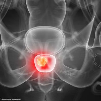
At the New York GU 14th Annual Interdisciplinary Prostate Cancer Congress® and Other Genitourinary Malignancies, Phillip J. Koo, MD, highlighted the potential of next-generation imaging to revolutionize the management of patients with prostate cancer.
Advertisement
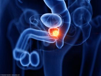
At the New York GU 14th Annual Interdisciplinary Prostate Cancer Congress® and Other Genitourinary Malignancies, Phillip J. Koo, MD, highlighted the potential of next-generation imaging to revolutionize the management of patients with prostate cancer.1
Koo, chief of Diagnostic Imaging, Northwest Region Oncology Physician Executive, Banner MD Anderson Cancer Center, Phoenix, Arizona, discussed the emergence of next-generation imaging; novel imaging agents, such as those targeting PSMA; and how these novel imaging techniques are “here to stay” in the prostate cancer paradigm.
Advantages of next-generation imaging
While conventional imaging techniques—such as bone scans, CT, or MRI—date back to the 1970s, their use does have limitations. For example, Koo noted that although bone scans are widely available and can help to identify bone lesions, they rarely identify asymptomatic disease or positively identify disease with the absence of a high PSA. Similarly, while CT scans can follow treatment response and can detect metastases, they cannot detect recurrent tumors and are dependent on size for nodal evaluation, which can result in poor sensitivity.
Therefore, this is why there has been a shift in focus toward next-generation imaging. In particular, this strategy may allow clinicians to see things that they wouldn’t be able to see using conventional imaging.
“It’s almost like the difference between a high-definition TV versus your standard TV,” Koo explained. “And we all recognize [that] with high-def, it’s just amazing what more you can see and how better appreciated that becomes.”
Moreover, he added, several specialized imaging techniques have emerged that may permit the acquisition of digital images at high resolution, which may have diagnostic potential to introduce new opportunities to more accurately assess disease burden. However, there is a lack of standardization or practical application of next-generation imaging in prostate cancer.
Next-generation imaging works by taking advantage of the unique biological aspects of prostate cancer carcinogens. Molecular imaging probes, of which several are already in clinical use or under evaluation, can be divided into images with increased cell metabolism, those that target prostate cancer–specific membrane proteins and receptor molecules, and those that bind to the bone matrix adjacent to metastases to the bone. Therefore, increased metabolism and vascular changes in prostate cancer cells can be evaluated with radiolabled analogs of choline, acetate, glucose, amino acids, and nucleotides, Koo explained.
Types of next-generation imaging
One example Koo provided of next-generation imaging was 18F-fluciclovine. Approved by the FDA in 2016, 18F-fluciclovine is a synthetic amino acid PET imaging agent that has demonstrated the ability to recognize amino acid transporters, which have been shown to be upregulated in many cancer cells, he explained. Additionally, because the radiotracer was not metabolized or incorporated into newly synthesized proteins, it may not interfere with normal protein synthesis.
“When the data were first reported in patients with biochemical recurrence with a PSA level of less than or equal to 0.79, it actually had a detection rate of 41% which is pretty remarkable given that lower PSA level. That’s why it created a lot of excitement, that we could image disease better at biochemical recurrence,” Koo said.2
Next, clinicians have moved into the evaluation of PSMA PET technology. PSMA is a membrane protein that has been shown to have a significant overexpression in prostatic tissue but low expression in normal tissue. Koo explained that high image quality could be achieved with PSMA PET because of the uptake in ligand bonding PSMA into the prostate cancer tumor cells. PSMA PET agents that are currently under investigation include 68Ga PSMA and 18F-DCFPyL PSMA; however, of note, neither imaging agent is FDA approved yet.
Next-generation imaging versus conventional imaging
In a prospective, single-center, open-label, single-arm comparative study, Michael S. Hoffman, MD, and colleagues compared 18F-fluciclovine and PSMA PET-CT scans for localizing biochemical recurrence of prostate cancer after radical prostatectomy in patients with low PSA concentrations (<2.0 ng/mL).
The investigators found that, among the 50 participants, detection rates were significantly lower with 18F-fluciclovine PET-CT (n = 13 [26%]; 95% CI, 15%-40%) than with PSMA PET-CT (n = 28 [56%]; 95% CI, 41%-70%), with an odds ratio (OR) of 4.8 (95% CI, 1.6-19.2; P = .0026) at the patient level. They concluded that PSMA should be the PET tracer of choice when PET-CT imaging is considered for subsequent treatment management decisions in patients with prostate cancer and biochemical recurrence after radical prostatectomy and low PSA concentrations.3
“The accuracy was much better than conventional imaging, [which was] not surprising, and they had a greater treatment impact,” Koo said. “It also had fewer uncertain results. So that’s something that I think is very important: An imaging test having less equivocal results is more powerful…hopefully this can get rid of that hedge in the future for radiology and nuclear medicine.”
Translating next-generation imaging into practice
With all of these data, Koo highlighted that it led to a “monumental day” in December 2020: The FDA approved the first PSMA-targeted PET imaging drug for men with prostate cancer.
“[The approval] actually had 2 indications. Number 1, it had the indications for use in patients with biochemical recurrence, which is something that we were used to. But the biggest difference for this approval was it was actually approved for patients at initial diagnosis,” Koo noted. “So it says that it’s indicated for suspected prostate cancer metastases, which are potentially curable by surgery or radiation therapy. This was pre-definitive treatment, which was something new for these next-generation imaging agents.”
Before concluding, Koo did note that next-generation imaging does no replace pathology: “PET CT can’t detect that microscopic level of detail disease in these patients, so we need to sort of separate the 2, because I often see that being assumed when it comes to pet imaging.”
However, Koo added, next generation imaging is here to stay. “For those of you in the audience who are resistant, I recommend that you start getting a bit more and more comfortable each year because it’s not going anywhere.”
References
1. Koo PJ. Next Generation Imaging in Prostate Cancer. Presented at: 14th Annual New York GU Congress; March 12-13, 2021.
2. Bach-Gansmo T, Nanni C, Nieh PT, et al. Multisite Experience of the Safety, Detection Rate and Diagnostic Performance of Fluciclovine (18 F) Positron Emission Tomography/Computerized Tomography Imaging in the Staging of Biochemically Recurrent Prostate Cancer. J Urol. 2017;197(3 Pt 1):676-638. doi:10.1016/j.juro.2016.09.117.
3. Calais J, Ceci F, Eiber M, et al. 8F-fluciclovine PET-CT and 68Ga-PSMA-11 PET-CT in patients with early biochemical recurrence after prostatectomy: a prospective, single-centre, single-arm, comparative imaging trial. Lancet Oncol. 2019;20(9):1286-1294. doi: 10.1016/S1470-2045(19)30415-2
Newsletter
Stay current with the latest urology news and practice-changing insights — sign up now for the essential updates every urologist needs.
Advertisement
Related Content
Advertisement


Latest CME


Advertisement
Advertisement
Trending on Urology Times
1
FDA approves sildenafil oral film for men with erectile dysfunction
2
Integration of doublet and triplet therapy in metastatic prostate cancer
3
The UroOnc Minute: Adjuvant Therapy in Renal Cell Carcinoma, with Brian Shuch, MD
4
URO-1 prostate biopsy devices adopted across Novant Health System as clinical study continues
5