
|Articles|November 24, 2020
Urology Times Urologists in Cancer Care
- UCC December 2020
- Volume 09
- Issue 04
Transurethral resection of the bladder tumor: Standard technique and new advancements
Author(s)Daniel D. Joyce, MD
Improvement in imaging modalities may help in performance of transurethral resection of the bladder tumor.
Advertisement
Since it was first described in 1910, transurethral resection of bladder tumor (TURBT) has evolved into the cornerstone of bladder cancer diagnosis and staging, and is one of the most common surgeries urologists perform. Following the introduction of video endoscopy in the late 1970s, TURBT has been characterized as a relatively simple procedure, often performed by junior residents in the academic setting. More recent data emphasize the importance of high-quality TURBT by experienced surgeons in order to improve patient outcomes.1 Significant variation in recurrence rates among urologists has been described, supporting the assumption that experienced technical skill matters. At our institution, junior residents slowly gain more responsibility as their technical skills develop under the close supervision of an experienced surgeon with a low threshold to take over the case when accurate staging may be difficult to obtain. Below, we discuss our standard technique for TURBT and describe new advancements that may help standardize outcomes in a procedure that has experienced little evolution during the past century.
TURBT technique
Preoperative considerations. Before operative intervention, a thorough history and physical should be performed. When possible, slides from prior resections are reviewed by our genitourinary specialized pathologists to ensure accurate staging and identify any variant histology. Routine lab work to assess renal function and rule out infection are obtained at the initial consultation, and imaging studies evaluating the upper urinary tract are performed before or at the time of TURBT. For patients with a history of non–muscle-invasive bladder cancer (NMIBC), we will often utilize blue light cystoscopy given the observed increased cancer detection (especially for carcinoma in situ [CIS]), decreased recurrence, and possibly decreased progression associated with its use. In addition, for those patients with discordant positive cytology and negative office cystoscopy, we will routinely use blue light technology in accordance with the American Urological Association (AUA) NMIBC guidelines. Although not yet incorporated into our practice, clinic blue light capabilities may further enhance patient care by increasing detection rates and avoiding unnecessary use of operative resources in patients with negative surveillance cystoscopy.
Procedure. Before and following resection, a bimanual exam is performed under sedation to assess clinical staging (fixation of bladder or palpable masses). The largest rigid resectoscope (ideally a 26 F with inflow and outflow ports to allow continuous irrigation) that the urethra will accommodate, with a 30° lens, is then introduced into the bladder. When indicated, a urine cytology is obtained. This can be performed by collecting the first emptied urine from the bladder (essentially a voided urine cytology) or as a formal bladder washing using saline. A careful white light cystourethroscopy is performed. In rare cases, a 70° lens may be needed to completely visualize the bladder. If enhanced cystoscopy techniques are planned, a repeat evaluation of the bladder is then performed using this technology. Obtaining photos of tumors can be useful for patient counseling and providing more detailed information for health care providers who are not present during the resection. Assessment of the size, location, grade, and presumed depth of invasion will inform the operative plan.
Tumor size. Smaller tumors in patients with thin bladder walls may be managed with cold cup biopsy, thereby mitigating the risk of bladder perforation. Alternatively, cautery loop resection can be performed, often en bloc, with one swipe. Larger tumors require a fragmented approach with successive loop resection until the base is exposed.
Tumor location. When resecting tumors in difficult locations where bladder injury is more likely, we will often use a staccato energy approach, which provides greater control and helps limit thermal injury. Tumors at the bladder dome and anterior wall may require abdominal pressure with the non-resecting hand to bring the mass into view. Occasionally, a long resectoscope may be required to reach the area of interest. The obturator reflex may be invoked when resecting lateral wall tumors, resulting in adduction of the ipsilateral leg and possible bladder perforation. Avoiding overdistention, use of bipolar energy source, resecting with decreased energy settings, and anesthetic paralysis may help limit this occurrence. For posterior wall tumors, bending the resection loop out to assimilate the contour of the bladder wall can provide safer and cleaner resection. For tumors near or overlying the ureteral orifices, pure cutting current should be used to avoid obstruction. The decision to leave a ureteral stent in this setting is surgeon dependent; however, existing data suggest that postresection hydronephrosis is rare regardless of stent use.2 Diverticular tumor resection carries a greater risk of bladder perforation given the lack of underlying detrusor muscle. When invasion is suspected, bladder perforation may be required in order to obtain adequate staging. In such cases, postoperative indwelling catheter placement should be utilized to allow adequate healing and avoid further complications.
Grade and depth of invasion. Low-grade tumors are associated with a low risk of progression and are increasingly being safely observed.3, 4 Smaller low-grade tumors can often be managed without anesthesia in an office setting using cautery or laser ablation following intravesical instillation of a lidocaine/bicarbonate solution. For high-grade tumors, obtaining detrusor muscle in the specimen is critical for staging purposes. Loop resection is performed to expose the tumor base. Further loop resection is then performed to obtain muscle. Alternatively, cold cup biopsy of the resection base is used in instances where bladder perforation is a concern.
Bipolar vs monopolar energy source. Bipolar energy, originally developed for transurethral resection of the prostate, is commonly used in TURBT as well, because of several potential advantages. Saline irrigation is used, diminishing the risk of TUR syndrome, which can occur after more extensive resections with water. Monopolar energy travels from the resection electrode through the body to the electrode attached to the patient’s skin, whereas bipolar energy travels between electrodes on the resection device. For lateral tumors where an obturator reflex is likely, bipolar may offer some advantage and limit bladder perforation. The difference in energy propagation may also limit cautery artifact in pathology samples. Finally, bipolar may offer more safety for patients with pacemakers and for pregnant women.5 Despite these theoretical advantages, comparative data are conflicting. In a recent systematic review of 13 randomized control trials comparing monopolar and bipolar TURBT, there was no observed difference in operative time, recurrence rate, bladder perforation, thermal damage, or overall complications.6
Postoperative considerations. Perioperative intravesical chemotherapy has been shown to reduce recurrence, especially in low-grade solitary tumors. Although data are more robust for the use of mitomycin C (MMC), we will often instill gemcitabine through an indwelling catheter in the operating room following resection of presumed low-grade tumors given the favorable adverse effect profile and recent phase 3 randomized control trial showing a recurrence rate reduction similar to that of MMC.7 Intravesical chemotherapy should be avoided following extensive resections or if bladder perforation is suspected.
Although postoperative continuous irrigation has been associated with lower recurrence rates, we typically avoid this in our patients because of concern for inducing or worsening bladder perforation.8
For high-grade T1 tumors, repeat resection is planned within 6 weeks to ensure proper staging and complete resection. Repeat resection is also considered for high-grade Ta tumors without muscle in the specimen or if incomplete resection is suspected.
Advancements in TURBT
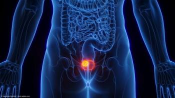
En bloc resection. En bloc resection (EBR) of bladder tumors has gained interest to avoid theoretical cancer cell circulation and out-of-field recurrences associated with traditional TURBT. Additionally, pathologic evaluation may be more accurate if tumor samples are kept intact. Advantages of this technique include improving muscle sampling and providing better hemostasis. A systematic review of EBR found a 95% rate of detrusor muscle inclusion in the specimen. Interestingly, this higher quality resection did not lead to decreased complication or recurrence rates.9 Use of electric energy, water dissection, holmium :YAG laser, thulium laser, and green-light laser have all been described.10 New endoscopic robotic instruments, such as those being developed by Virtuoso Surgical (Figure), provide a more user-friendly interface for EBR, which may translate into greater consistency in TURBT quality, regardless of skill level. Still, endoscopic removal of intact tumors greater than 3 cm remains a challenge. Various methods have been proposed to address this, including the use of laparoscopic forceps, nephroscopy sheaths, nylon mesh retrieval nets, and endo-bags (such as those used in GI endoscopy).11-13 More data are needed to justify EBR adoption into common practice.
Bladder imaging. Improved imaging modalities may offer further consistency and accuracy in clinical staging of bladder cancer. Knowing the depth of invasion before resection may lead to reducing the observed 50% residual tumor rate and 10% progression rate found at repeat resection through better operative planning and higher-precision resection. Intraoperative use of optical coherence tomography and confocal laser endomicroscopy are emerging technologies that may improve diagnostic accuracy; however, further investigation is required to clarify user variability and its impact on oncologic outcomes.14 Similarly, preoperative use of multiparametric magnetic resonance imaging (MRI) and development of the Vesical Imaging-Reporting and Data System (VI-RADS) score appear to improve clinical staging of muscle-invasive disease.15 Similar to its increasing popularity in prostate cancer diagnosis, MRI may become more common for bladder cancer staging as radiology interpretation becomes more consistent and standardized.
Future directions. Artificial intelligence and machine learning offer the promise of standardized diagnostic accuracy and improved cancer detection.16, 17 Digital bladder mapping software is being developed to reduce cystoscopy interobserver variability.18 Ultimately, technology that increases detection of bladder cancer and decreases TURBT quality variability may drastically change a procedure that has seen little evolution over the past 100 years.
Joyce is a urology resident at Vanderbilt University Medical Center, Nashville, Tennessee and future urologic oncology fellow at the Mayo Clinic in Rochester, Minnesota.
References
1. Mariappan P, Finney SM, Head E, et al; Edinburgh Urological Cancer Group. Good quality white-light transurethral resection of bladder tumours (GQ-WLTURBT) with experienced surgeons performing complete resections and obtaining detrusor muscle reduces early recurrence in new non-muscle-invasive bladder cancer: validation across time and place and recommendation for benchmarking. BJU Int. 2012;109(11):1666-1673. doi:10.1111/j.1464-410X.2011.10571.x
2. Mano R, Shoshany O, Baniel J, Yossepowitch, O. Resection of ureteral orifice during transurethral resection of bladder tumor: functional and oncologic implications. J Urol. 2012;188(6):2129-2133. doi:10.1016/j.juro.2012.08.006
3. Soloway MS, Bruck DS, Kim SS. Expectant management of small, recurrent, noninvasive papillary bladder tumors. J Urol. 2003;170(2 Pt 1):438-441. doi:10.1097/01.ju.0000076621.71247.6c
4. Pruthi RS, Baldwin N, Bhalani V, Wallen EM. Conservative management of low risk superficial bladder tumors. J Urol. 2008;179(1):87-90. doi:10.1016/j.juro.2007.08.171
5. Zhao C, Tang K, Yang H, Xia D, Chen Z. Bipolar versus monopolar transurethral resection of nonmuscle-invasive bladder cancer: a meta-analysis. J Endourol. 2016;30(1):5-12. doi:10.1089/end.2015.0410
6. Xie K, Cao D, Wei Q, et al. Bipolar versus monopolar transurethral resection of non-muscle-invasive bladder cancer: a systematic review and meta-analysis of randomized controlled trials. World J Urol. Published online May 27, 2020. doi:10.1007/s00345-020-03271-3
7. Messing EM, Tangen CM, Lerner SP, et al. Effect of intravesical instillation of gemcitabine vs saline immediately following resection of suspected low-grade non–muscle-invasive bladder cancer on tumor recurrence: SWOG S0337 randomized clinical trial. JAMA. 2018;319(18):1880-1888. doi:10.1001/jama.2018.4657
8. Zhou Z, Zhao S, Lu Y, et al. Meta-analysis of efficacy and safety of continuous saline bladder irrigation compared with intravesical chemotherapy after transurethral resection of bladder tumors. World J Urol. 2019;37(6):1075-1084. doi:10.1007/s00345-019-02628-7
9. Kramer MW, Altieri V, Hurle R, et al. Current evidence of transurethral en-bloc resection of nonmuscle invasive bladder cancer. Eur Urol Focus. 2017;3(6):567-576. doi:10.1016/j.euf.2016.12.004
10. Pastuszak A, Zdrojowy R, Poletajew S, Adamowicz J, Krajewski W. Technical developments in transurethral resection of bladder tumours. Contemp Oncol (Pozn). 2019;23(4):195-201. doi:10.5114/wo.2019.91530
11. Fritsche HM, Otto W, Eder F, et al. Water-jet-aided transurethral dissection of urothelial carcinoma: a prospective clinical study. J Endourol. 2011;25(10):1599-1603. doi:10.1089/end.2011.0042
12. Maurice MJ, Vricella GJ, MacLennan G, Buehner P, Ponsky LE. Endoscopic snare resection of bladder tumors: evaluation of an alternative technique for bladder tumor resection. J Endourol. 2012;26(6):614-617. doi:10.1089/end.2011.0587.
13. Naselli A, Introini C, Germinale F, Spina B, Puppo P. En bloc transurethral resection of bladder lesions: a trick to retrieve specimens up to 4.5 cm. BJU Int. 2012;109(6):960-963. doi:10.1111/j.1464-410X.2012.10982.x
14. Tully K, Palisaar RJ, Brock M, et al. Transurethral resection of bladder tumours: established and new methods of tumour visualisation. Transl Androl Urol. 2019;8(1):25-33. doi:10.21037/tau.2018.12.12
15. Carando R, Afferi L, Marra G, et al. The effectiveness of multiparametric magnetic resonance imaging in bladder cancer (Vesical Imaging-Reporting and Data System): a systematic review. Arab J Urol. 2020;18(2):67-71. doi:10.1080/2090598X.2020.1733818
16. Thompson RF, Valdes G, Fuller CD, et al. Artificial intelligence in radiation oncology: a specialty-wide disruptive transformation? Radiother Oncol. 2018;129(3):421-426. doi:10.1016/j.radonc.2018.05.030
17. Hosny A, Parmar C, Quackenbush J, Schwartz LH, Aerts HJWL. Artificial intelligence in radiology. Nat Rev Cancer. 2018;18(8):500-510. doi:10.1038/s41568-018-0016-5
18. Kriegmair MC, Bergen T, Ritter M, et al. Digital mapping of the urinary bladder: potential for standardized cystoscopy reports. Urology. 2017;104:235-241. doi:10.1016/j.urology.2017.02.019
Articles in this issue
over 5 years ago
PARP inhibitors: Treating mCRPC from a genetic basisover 5 years ago
The promise of precision medicine for urologic cancersover 5 years ago
Radical nephrectomy before or after systemic therapy for mRCCover 5 years ago
Urologic implications of Lynch syndromeAdvertisement
Related Content
Advertisement


Latest CME


Advertisement
Advertisement
Trending on Urology Times
1
The UroOnc Minute: AUA 2026 Update on Prostate Cancer Early Detection, With Badrinath R. Konety, MBBS
2
FDA approves enfortumab vedotin plus pembrolizumab for MIBC
3
Daniel George, MD, on the significance of the capivasertib approval for PTEN-deficient prostate cancer
4
EV plus pembro approval reframes first conversation in MIBC diagnosis
5